


Contrast-Enhanced 2D-3D Mammography in Oklahoma City

Next-Level Clarity for Complex Breast Imaging
When standard mammography leaves questions unanswered, Contrast-Enhanced 2D-3D Mammography (CEM) delivers greater clarity. This advanced imaging technique highlights areas of abnormal blood flow, making it easier to detect and evaluate potential concerns, especially in dense breast tissue or high-risk patients.

How Contrast-Enhanced Mammography Works
A small IV is used to administer an iodine-based contrast agent shortly before imaging. The exam combines standard mammography images with information about contrast uptake in the breast, allowing areas with increased blood flow to stand out more clearly.
What’s the Difference Between 2D and 3D Mammography?
Understanding your imaging options can help you feel more confident in your care. Here’s how 2D and 3D mammography differ:
2D Mammography:
Captures flat images of the breast from two angles, top-to-bottom and side-to-side. It’s fast, effective, and commonly used for routine screenings. However, overlapping breast tissue can sometimes hide abnormalities or create false alarms.
3D Mammography (Tomosynthesis)
Takes multiple images of the breast from different angles and reconstructs them into thin, detailed layers. This “slice-by-slice” view reduces the chances of missing small cancers and cuts down on unnecessary callbacks by giving radiologists a clearer picture. This is especially helpful in women with dense breast tissue.
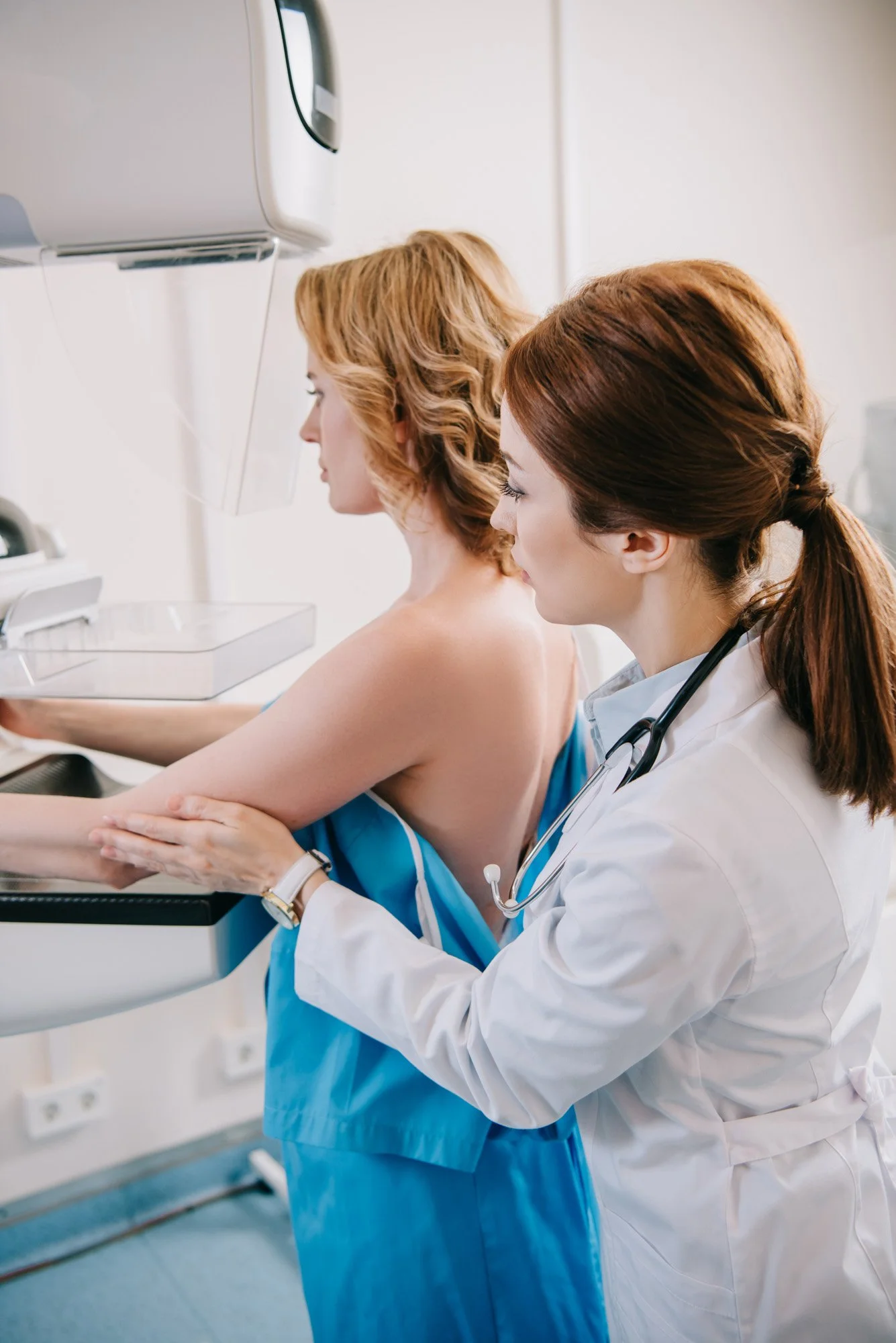
Why Choose CEM Over Standard Imaging?
-
CEM can provide additional information when an area is difficult to evaluate on standard mammography alone.
-
The exam provides detailed mammography images while also showing contrast uptake in a single visit.
-
For selected patients, CEM may be considered when MRI is not practical because of certain implants, claustrophobia or other medical considerations.

When CEM May Be Recommended
-
Dense breast tissue or an inconclusive mammogram result
-
Evaluation of a newly diagnosed breast cancer
-
Supplemental imaging for some higher-risk patients
-
When breast MRI is not an appropriate or practical option
What to Expect from a Contrast -Enhanced Mammography Exam
A small IV is placed for the contrast agent, followed by a brief waiting period before mammogram images are taken. The full visit generally takes about 20 to 30 minutes. Our team reviews relevant allergies and health history beforehand, explains each step and works to keep you comfortable throughout the exam.
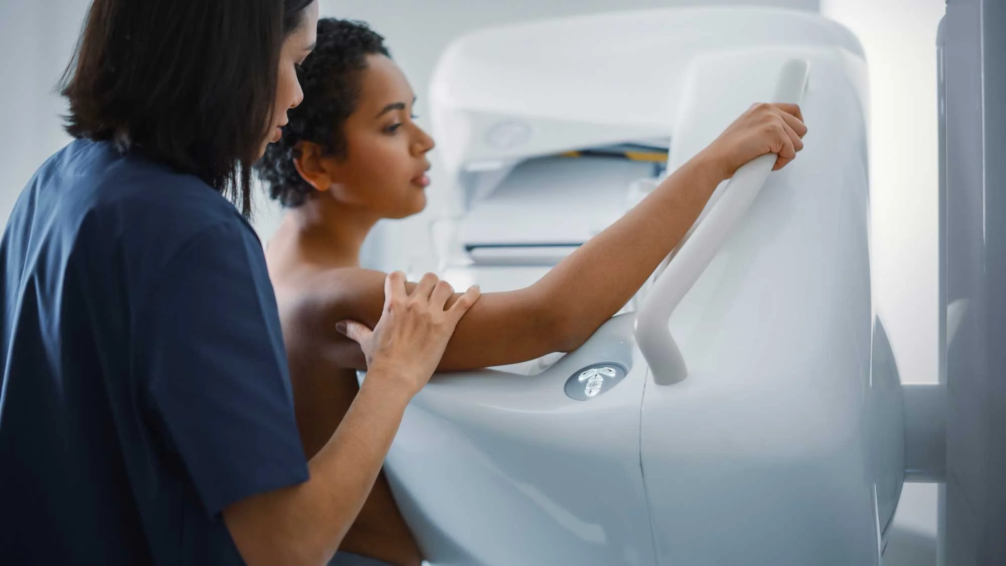
A Closer Look at Your Mammogram Experience
This video gives you a closer look at our mammogram services. You'll learn what to expect, how we care for you, and why early detection matters.